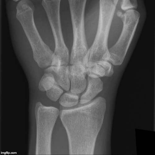
Tunnel View Wrist X Ray . The view can be used to visualize hook of hamate, trapezium, and pisiform fractures. the beam is aimed through the carpal tunnel. The image displays the inner structure. “carpal tunnel” view when: Consider this view when a patient has pain at hypothenar eminence or a sensory deficit in the ulnar distribution.
from www.aliem.com
“carpal tunnel” view when: the beam is aimed through the carpal tunnel. Consider this view when a patient has pain at hypothenar eminence or a sensory deficit in the ulnar distribution. The image displays the inner structure. The view can be used to visualize hook of hamate, trapezium, and pisiform fractures.
EMRad Radiologic Approach to the Traumatic Wrist
Tunnel View Wrist X Ray “carpal tunnel” view when: “carpal tunnel” view when: The view can be used to visualize hook of hamate, trapezium, and pisiform fractures. the beam is aimed through the carpal tunnel. Consider this view when a patient has pain at hypothenar eminence or a sensory deficit in the ulnar distribution. The image displays the inner structure.
From www.wikiradiography.net
Wrist Radiographic Anatomy wikiRadiography Tunnel View Wrist X Ray Consider this view when a patient has pain at hypothenar eminence or a sensory deficit in the ulnar distribution. The image displays the inner structure. “carpal tunnel” view when: the beam is aimed through the carpal tunnel. The view can be used to visualize hook of hamate, trapezium, and pisiform fractures. Tunnel View Wrist X Ray.
From www.youtube.com
Wrist Fracture Xrays YouTube Tunnel View Wrist X Ray The view can be used to visualize hook of hamate, trapezium, and pisiform fractures. the beam is aimed through the carpal tunnel. The image displays the inner structure. Consider this view when a patient has pain at hypothenar eminence or a sensory deficit in the ulnar distribution. “carpal tunnel” view when: Tunnel View Wrist X Ray.
From radiopaedia.org
Image Tunnel View Wrist X Ray The image displays the inner structure. the beam is aimed through the carpal tunnel. The view can be used to visualize hook of hamate, trapezium, and pisiform fractures. Consider this view when a patient has pain at hypothenar eminence or a sensory deficit in the ulnar distribution. “carpal tunnel” view when: Tunnel View Wrist X Ray.
From geekymedics.com
Wrist Xray Interpretation OSCE Guide Geeky Medics Tunnel View Wrist X Ray The view can be used to visualize hook of hamate, trapezium, and pisiform fractures. Consider this view when a patient has pain at hypothenar eminence or a sensory deficit in the ulnar distribution. the beam is aimed through the carpal tunnel. “carpal tunnel” view when: The image displays the inner structure. Tunnel View Wrist X Ray.
From www.mdpi.com
JCM Free FullText The Radiographic Risk Factors Predicting Occurrence of Idiopathic Carpal Tunnel View Wrist X Ray The view can be used to visualize hook of hamate, trapezium, and pisiform fractures. the beam is aimed through the carpal tunnel. “carpal tunnel” view when: Consider this view when a patient has pain at hypothenar eminence or a sensory deficit in the ulnar distribution. The image displays the inner structure. Tunnel View Wrist X Ray.
From www.researchgate.net
Xray image showing the left hand wrist in dorsal view. The carpal... Download Scientific Diagram Tunnel View Wrist X Ray The image displays the inner structure. the beam is aimed through the carpal tunnel. “carpal tunnel” view when: The view can be used to visualize hook of hamate, trapezium, and pisiform fractures. Consider this view when a patient has pain at hypothenar eminence or a sensory deficit in the ulnar distribution. Tunnel View Wrist X Ray.
From www.bmj.com
The carpal bones on a lateral plain radiograph of the wrist The BMJ Tunnel View Wrist X Ray the beam is aimed through the carpal tunnel. The image displays the inner structure. “carpal tunnel” view when: The view can be used to visualize hook of hamate, trapezium, and pisiform fractures. Consider this view when a patient has pain at hypothenar eminence or a sensory deficit in the ulnar distribution. Tunnel View Wrist X Ray.
From www.youtube.com
Normal wrist xray radiographic anatomy tutorial YouTube Tunnel View Wrist X Ray the beam is aimed through the carpal tunnel. Consider this view when a patient has pain at hypothenar eminence or a sensory deficit in the ulnar distribution. “carpal tunnel” view when: The view can be used to visualize hook of hamate, trapezium, and pisiform fractures. The image displays the inner structure. Tunnel View Wrist X Ray.
From orthoinfo.aaos.org
Arthritis of the Wrist OrthoInfo AAOS Tunnel View Wrist X Ray “carpal tunnel” view when: The view can be used to visualize hook of hamate, trapezium, and pisiform fractures. the beam is aimed through the carpal tunnel. The image displays the inner structure. Consider this view when a patient has pain at hypothenar eminence or a sensory deficit in the ulnar distribution. Tunnel View Wrist X Ray.
From ce4rt.com
CE4RT Radiographic Positioning of the Wrist for Xray Technologists Tunnel View Wrist X Ray the beam is aimed through the carpal tunnel. The view can be used to visualize hook of hamate, trapezium, and pisiform fractures. “carpal tunnel” view when: The image displays the inner structure. Consider this view when a patient has pain at hypothenar eminence or a sensory deficit in the ulnar distribution. Tunnel View Wrist X Ray.
From www.cortho.org
Causes and Management of Wrist Joint Pain Complete Orthopedics Tunnel View Wrist X Ray The image displays the inner structure. “carpal tunnel” view when: the beam is aimed through the carpal tunnel. The view can be used to visualize hook of hamate, trapezium, and pisiform fractures. Consider this view when a patient has pain at hypothenar eminence or a sensory deficit in the ulnar distribution. Tunnel View Wrist X Ray.
From www.wikiradiography.net
Carpal Tunnel Radiography wikiRadiography Tunnel View Wrist X Ray The image displays the inner structure. “carpal tunnel” view when: The view can be used to visualize hook of hamate, trapezium, and pisiform fractures. Consider this view when a patient has pain at hypothenar eminence or a sensory deficit in the ulnar distribution. the beam is aimed through the carpal tunnel. Tunnel View Wrist X Ray.
From www.bmj.com
Lateral radiograph of the right wrist The BMJ Tunnel View Wrist X Ray Consider this view when a patient has pain at hypothenar eminence or a sensory deficit in the ulnar distribution. the beam is aimed through the carpal tunnel. “carpal tunnel” view when: The view can be used to visualize hook of hamate, trapezium, and pisiform fractures. The image displays the inner structure. Tunnel View Wrist X Ray.
From www.kenhub.com
Radiological anatomy Xray, CT, MRI Kenhub Tunnel View Wrist X Ray the beam is aimed through the carpal tunnel. Consider this view when a patient has pain at hypothenar eminence or a sensory deficit in the ulnar distribution. The view can be used to visualize hook of hamate, trapezium, and pisiform fractures. “carpal tunnel” view when: The image displays the inner structure. Tunnel View Wrist X Ray.
From www.animalia-life.club
X Ray Wrist Lateral View Tunnel View Wrist X Ray “carpal tunnel” view when: Consider this view when a patient has pain at hypothenar eminence or a sensory deficit in the ulnar distribution. the beam is aimed through the carpal tunnel. The view can be used to visualize hook of hamate, trapezium, and pisiform fractures. The image displays the inner structure. Tunnel View Wrist X Ray.
From ally.perka.org
Carpal Tunnel X Ray Positioning ally Tunnel View Wrist X Ray Consider this view when a patient has pain at hypothenar eminence or a sensory deficit in the ulnar distribution. The image displays the inner structure. the beam is aimed through the carpal tunnel. The view can be used to visualize hook of hamate, trapezium, and pisiform fractures. “carpal tunnel” view when: Tunnel View Wrist X Ray.
From www.wikiradiography.net
Carpal Tunnel Radiography wikiRadiography Tunnel View Wrist X Ray The image displays the inner structure. The view can be used to visualize hook of hamate, trapezium, and pisiform fractures. the beam is aimed through the carpal tunnel. Consider this view when a patient has pain at hypothenar eminence or a sensory deficit in the ulnar distribution. “carpal tunnel” view when: Tunnel View Wrist X Ray.
From bonepit.com
UCSD Musculoskeletal Radiology Tunnel View Wrist X Ray The view can be used to visualize hook of hamate, trapezium, and pisiform fractures. “carpal tunnel” view when: Consider this view when a patient has pain at hypothenar eminence or a sensory deficit in the ulnar distribution. The image displays the inner structure. the beam is aimed through the carpal tunnel. Tunnel View Wrist X Ray.